
Every 2 seconds someone in the world will have a stroke for the first time. There were almost 17 million incidences of first-time stroke worldwide in 2010. Stroke is one of the leading causes of long-term adult disability.
Are you aware of the impact of post-stroke aphasia?

Post-stroke aphasia accounts for around 85% of all cases of aphasia, is present in 21-38% of post-stroke patients (Laska et al., 2001; Berthier, 2005), and poses a major challenge in neurorehabilitation.
While spontaneous post-stroke aphasia recovery occurs, this largely takes place in the first 2 to 3 months after a stroke with a slower rate and longer progress time compared with spontaneous motor recovery (Sarno and Levita, 1981; Wade et al., 1986). Further, 12% of post-stroke survivors are left with some degree of chronic communication deficit even after vigorous treatment (Wade et al., 1986; Lazar et al., 2010).
Patients with post-stroke aphasia experience longer length of stays, greater morbidity, and greater mortality than those without aphasia and therefore incur greater costs (Ellis et al., 2012). Additionally, people with aphasia tend to participate in fewer activities and report worse quality of life after stroke than those without aphasia (Hilari, 2011).
Speeding post-stroke language recovery – the real unmet need!
The aphasic population is heterogeneous, with individual profiles of language impairment varying in terms of severity and degree of involvement across the modalities of language processing, including the expression and comprehension of speech, reading, writing and gesture (Parr et al., 1997; Code and Herrmann, 2003).

Speech and language therapy (SLT) is the most commonly employed treatment in aphasia. Generally, SLT is tailored to meet the individual needs of patients. Nevertheless, its therapeutic effects are quite variable and usually modest (Brady et al., 2012).
The potential of neuromodulation in post-stroke language rehab
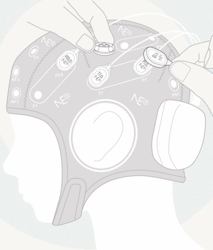
Neurorehabilitation with non-invasive brain stimulation techniques (NIBS) ‒ particularly repetitive transcranial magnetic stimulation (rTMS) or transcranial direct current stimulation (tDCS) ‒ may enhance the effects of SLT in selected patients. rTMS and tDCS, have shown promise as potential approaches for enhancing aphasia treatment due to the evidence of modulating neural reorganization after stroke.
Like rTMS, tDCS can alter cortical excitability in predictable ways. However, tDCS is characterized as neuromodulatory rather than neurostimulatory, since the currents delivered during tDCS are not sufficient to directly generate or inhibit action potentials. tDCS currents modulate neural resting membrane potentials, in which anodal tDCS (a-tDCS) increases cortical excitability and cathodal (c-tDCS) decreases cortical excitability (Nitsche and Paulus, 2000). tDCS can easily be administered during behavioral treatment, and is less expensive and likely to be better accepted by patients than rTMS (Floel et al., 2011; Floel, 2014). Nevertheless, implications for clinical practice should be ascertained in larger multicentre trials.
Neuroplasticity and the concept of interhemispheric inhibition

Language recovery after a stroke depends significantly on the degree of neuroplastic change, which is usually associated with reorganization and reconnection of the lesioned and perilesional dominant hemisphere regions, acquisition or unmasking of the homologous language area in the non-dominant hemisphere, or activation of the non-dominant cortical region (Hamilton et al., 2011).
As already analyzed by Shah et al. (2013), many studies employing tDCS as a therapy for aphasia have adopted approaches that are broadly consistent with an interhemispheric inhibition model of aphasia recovery. That is, a-tDCS investigations are mainly centered on left hemisphere language areas in order to increase the excitability in the perilesional and residual fronto-temporal areas (Baker et al., 2010; Fiori et al., 2011; Fridriksson et al., 2011; Marangolo et al., 2013), whereas c-tDCS is generally applied to the right homotopic areas to inhibit over activation (due to transcollasal disinhibition) in the contralesional right homologs. In a recent Cochrane meta-analysis, Elsner et al. (2013) evaluated five tDCS interventional trials with sham-controls involving 54 post-stroke aphasic patients. Although these studies using tDCS (a-tDCS or c-tDCS) in combination with SLT favored the intervention in each of these five trials (Monti et al., 2008; Floel et al., 2011; Kang et al., 2011; Marangolo et al., 2011; You et al. 2011), confidence interval width did not allow the results to be generalized. Elsner et al. (2013) did however state that when considering only c-tDCS over the non-lesioned hemisphere versus sham-tDCS, the effect on naming accuracy rises and the probability of error declines.
Further steps towards enhancing our quality-of-life….
Otal et al. (2015) identified and summarized RCTs and randomized controlled cross-over trials assessing the clinical efficacy of NIBS techniques in their inhibitory form (i.e., low-frequency rTMS or cathodal tDCS) over the unaffected non-language dominant hemisphere as an adjunct to SLT for post-stroke aphasia rehabilitation. When outcome measures were considered comparable, the authors combined these in an exploratory meta-analysis. This study allowed to specifically examine the neuroplastic process underlying aphasia recovery in adults considering the concept of reducing interhemispheric competition. The results reflect that low-frequency rTMS and c-tDCS over the unaffected non-language dominant hemisphere may be a promising approach compatible with the concept of interhemispheric inhibition (for specific meta-analysis details refer to the original article by clicking on Frontiers in Human Neuroscience).
Take home message
Neromodulation is promising!, yet, further multicenter RCTs with larger populations and homogenous intervention protocols are required to confirm these and the longer-term effects of rTMS and tDCS in post-stroke aphasia rehabilitation.
Baker JM, Rorden C, Fridriksson J. Using transcranial direct-current stimulation to treat stroke patients with aphasia. Stroke (2010) 41:1229-1236. doi: 10.1161/STROKEAHA.109.576785
Berthier ML. Postroke aphasia – epidemiology, pathophysiology and treatment. Drugs Aging (2005) 22:163-182. doi:10.2165/00002512-200522020-00006
Brady MC, Kelly H, Godwin J, Enderby P. Speech and language therapy for aphasia following stroke. Cochrane Database Syst. Rev.(2012) 5:CD000425. doi: 10.1002/14651858.CD000425
Code C, Herrmann M. The relevance of emotional and psychosocial factors in aphasia to rehabilitation. Neuropsychol Rehabil (2003) 13(1-2):109-32. doi: 10.1080/09602010244000291
Ellis C, Simpson AN, Bonilha H, Mauldin PD, Simpson KN. The one-year attributable cost of poststroke aphasia. Stroke (2012) 43(5):1429-31. doi: 10.1161/STROKEAHA.111.647339
Elsner B, Kugler J, Pohl M, Mehrholz J. Transcranial direct current stimulation (tDCS) for improving aphasia in patients after stroke. Cochrane Database (2013). Syst Rev 6:CD009760. doi: 10.1002/14651858.CD009760
Fiori V, Coccia M, Marinelli CV, Vecchi V, Bonifazi S, Ceravolo MG, Provinciali L, Tomaiuolo F, Marangolo P. Transcranial direct current stimulation improves word retrieval in healthy and nonfluent aphasic subjects. J Cogn Neurosci (2011) 23(9):2309-23. doi: 10.1162/jocn.2010.21579.
Flöel A, Meinzer M, Kirstein R, Nijhof S, Deppe M, Knecht S, Breitenstein C. Short-term anomia training and electrical brain stimulation. Stroke (2011) 42(7): 2065-2067. doi: 10.1161/STROKEAHA.110.609032
Flöel A. tDCS-enhanced motor and cognitive function in neurological diseases. Neuroimage (2014) 85 Pt 3:934-47. doi: 10.1016/j.neuroimage.2013.05.098
Fridriksson J, Richardson JD, Baker JM, Rorden C. Transcranial direct current stimulation improves naming reaction time in fluent aphasia: A double-blind, sham-controlled study. Stroke (2011) 42(3):819-821. doi: 10.1161/STROKEAHA.110.600288
Hamilton RH, Chrysikou EG, Coslett B. Mechanisms of aphasia recovery after stroke and the role of noninvasive brain stimulation. Brain Lang (2011) 118:40-50. doi: 10.1016/j.bandl.2011.02.005
Hilari K. The impact of stroke: are people with aphasia different to those without? Disabil Rehabil (2011) 33(3):211-8. doi: 10.3109/09638288.2010.508829.
Kang EK, Kim YK, Sohn HM, Cohen LG, Paik NJ. Improved picture naming in aphasia patients treated with cathodal tDCS to inhibit the right Broca’s homologue area. Restor Neurol Neurosci (2011) 29(3):141-52. doi: 10.3233/RNN-2011-0587
Laska AC, Hellblom A, Murray V, Kahan T, von Arbin M. Aphasia in acute stroke and relation to outcome. J Intern Med (2001) 249(5):413-22.
Lazar RM, Minzer B, Antoniello D, Festa JR, Krakauer JW, Marshall RS. Improvement in aphasia scores after stroke is well predicted by initial severity. Stroke (2010) 41:1485-1488. doi: 10.1161/STROKEAHA
Marangolo P, Marinelli CV, Bonifazi S, Fiori V, Ceravolo MG, Provinciali L, Tomaiuolo F. Electrical stimulation over the left inferior frontal gyrus (IFG) determines long-term effects in the recovery of speech apraxia in three chronic aphasics. Behav Brain Res (2011) 225(2):498-504. doi: 10.1016/j.bbr.2011.08.008
Marangolo P, Fiori V, Calpagnano MA, Campana S, Razzano C, Caltagirone C, Marini A. tDCS over the left inferior frontal cortex improves speech production in aphasia. Front. Hum. Neurosci. (2013) 7:539. doi:10.3389/fnhum.2013.00539
Monti A, Cogiamanian F, Marceglia S, Ferrucci R, Mameli F, Mrakic-Sposta S, Vergari M, Zago S, Priori A. Improved naming after transcranial direct current stimulation in aphasia. J Neurol Neurosurg Psychiatry (2008)79(4):451-3.
Nitsche A, Paulus W. Excitability changes induced in the human motor cortex by weak transcranial direct current stimulation. J Physiol (2000) 527 Pt 3:633-9.
Otal B, Olma M, Flöel A, Wellwood I. “Inhibitory non-invasive brain stimulation to homologous language regions as an adjunct to speech and language therapy in post-stroke aphasia: a meta-analysis”, Frontiers in Human Neuroscience 04/2015; 9(236). doi:10.3389/fnhum.2015.00236
Parr S, Byng S, Gilpin S, Ireland C. Talking about Aphasia: Living with loss of language after stroke. Buckingham: OUP, (1997).
Sarno MT, Levita E. Some observations on the nature of recovery in global aphasia after stroke. Brain Lang (1981)13:1-12.
Shah PP, Szaflarski JP, Allendorfer J, Hamilton RH. Induction of neuroplasticity and recovery in post-stroke aphasia by non-invasive brain stimulation. Front Hum Neurosci (2013) 7:888. doi: 10.3389/fnhum.2013.00888
Stroke statistics, January 2015 – stroke.org.uk
Wade DT, Hewer RL, David RM, Enderby PM. Aphasia after stroke: natural history and associated deficits. J Neurol Neurosurg Psychiatry (1986) 49(1):11-16.
You DS, Kim DY, Chun MH, Jung SE, Park SJ. Cathodal transcranial direct current stimulation of the right Wernicke’s area improves comprehension in subacute stroke patients. Brain Lang (2011) 119(1):1-5. doi: 10.1016/j.bandl.2011.05.002
Part of this work is based on the original manuscript published by the same author on Frontiers in Human Neuroscience in April 2015, Inhibitory non-invasive brain stimulation to homologous language regions as an adjunct to speech and language therapy in post-stroke aphasia: a meta-analysis. The analysis was performed prior to joining the company (NE) without any conflict of interest. This text contains direct paragraphs from Otal et al. (2015) publication.